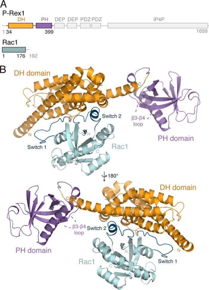
FIGURE 1.
Structure of the P-Rex1·Rac1 complex. A, schematic representation of the domain layout of P-Rex1, highlighting the position of the DH (yellow) and PH (purple) domains. Rac1 is shown in teal. B, the structure of P-Rex1-(34–399) bound to Rac1-(1–176) shown schematically. The position of the missing β3-β4 loop within the P-Rex1 PH domain is indicated.

