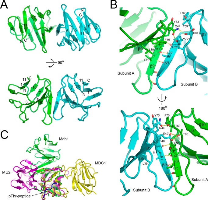
FIGURE 3.
Dimeric structure of Mdb1-FHA. A, ribbon representation of Mdb1-FHA dimer structure shown in two orthogonal views. Subunits A and B are colored green and cyan, respectively. The β-strands are numbered as in Fig. 2, and the N and C termini are labeled. B, the dimer interface viewed from two opposite sides. Residues involved in dimerization are shown as sticks with oxygen colored red and nitrogen colored blue. Hydrogen bonds are shown as yellow dashed lines. C, one subunit of the FHA dimers of Mdb1 (green), MU2 (magenta), and MDC1 (yellow) are aligned. The bound Thr(P)-peptide of MDC1 is shown in blue.

