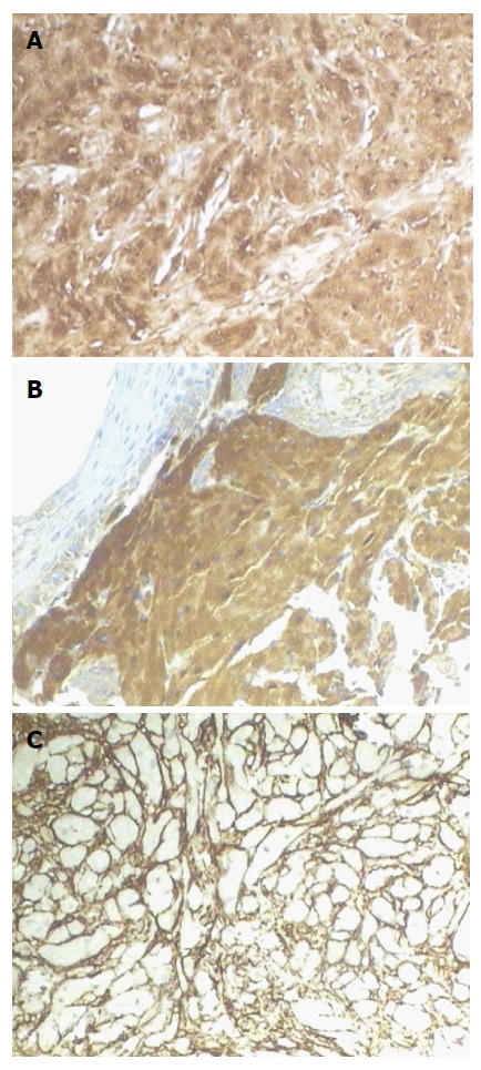
Figure 3.

Immunohistochemical and histochemical staining. Envision method, × 100. A: S-100 was strongly positive in tumor cells; B: CD68 was strongly positive in tumor cells; C: CD34 was negative in tumor cells, but the surrounding mesenchymal cells were positive for CD34.
