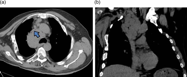
Figure 2.

A chest CT scan without intravenous contrast demonstrating (A) superior vena cava (indicated by a bold arrow) compressed by bulky, confluent mediastinal lymph nodes, (B) mediastinal lymphadenopathy extending into the supraclavicular region of the neck without evidence of a bronchial or parenchymal lung mass.
