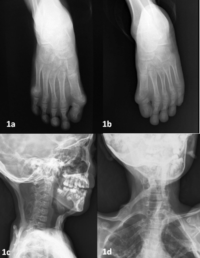
Figure 1.
(A, B) Plain radiographs bilateral feet showing congenital deformities of great toes because of small proximal phalanx and bilateral hallux valgus. (C, D) Plain radiographs neck anteroposterior (AP) and lateral view showing well-defined cord-like ossifications of anterior and posterior neck muscles with straightening of the cervical spine.

