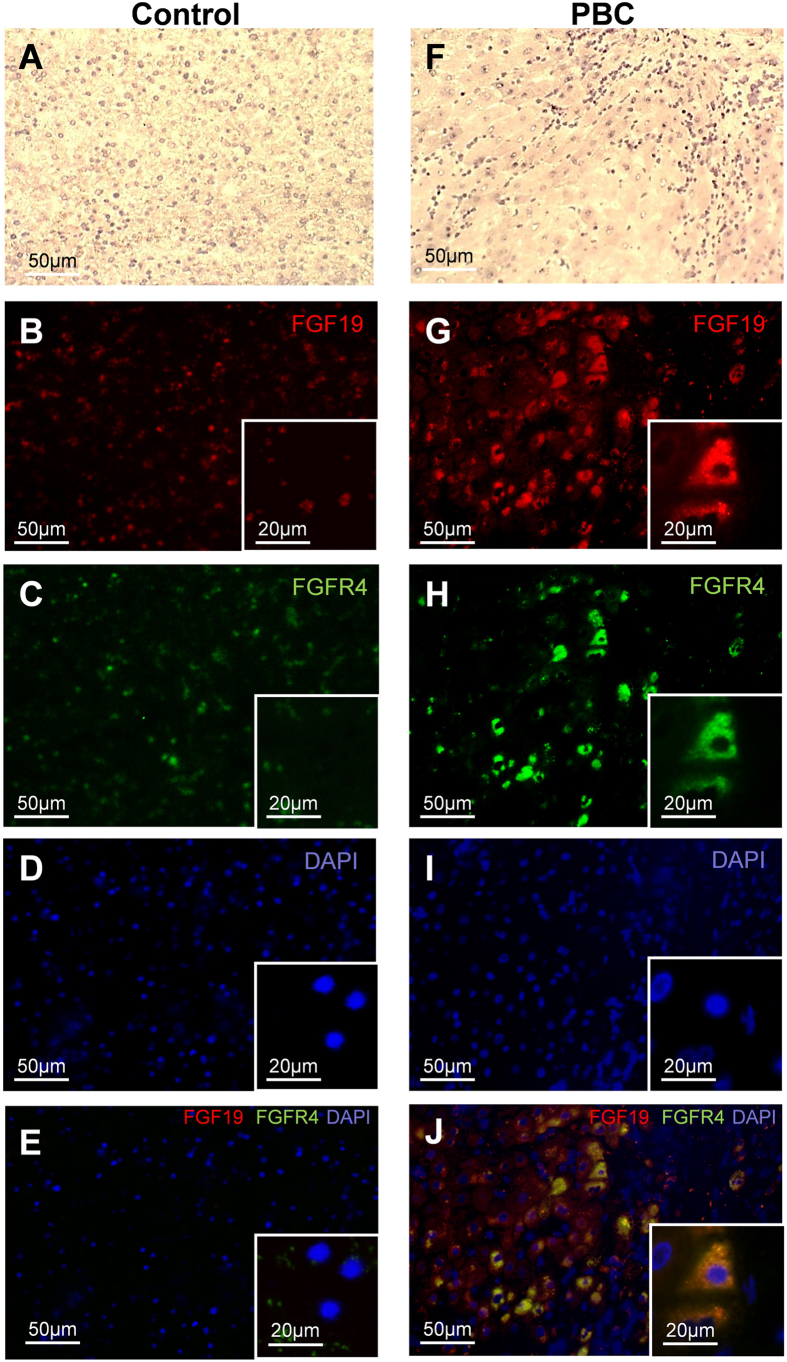
Figure 3. The hepatic expression of FGF19 and FGFR4 protein in cirrhotic patients with PBC and controls.
Representative light micrographs of hematoxylin stained liver sections of control (A) and PBC (F). Immunofluorescence staining of liver tissue showed that in comparison to controls (B,C) expression of both (G) FGF19 (red) and (H) FGFR4 (green) were substantively increased in PBC. Nuclei (blue) stained with DAPI (D,I). Merged immunofluorescent images of FGF19, FGFR4 and DAPI (E,J) demonstrated that in chronic cholestatic PBC livers FGF19 produced by hepatocytes binds to the FGFR4 (J).

