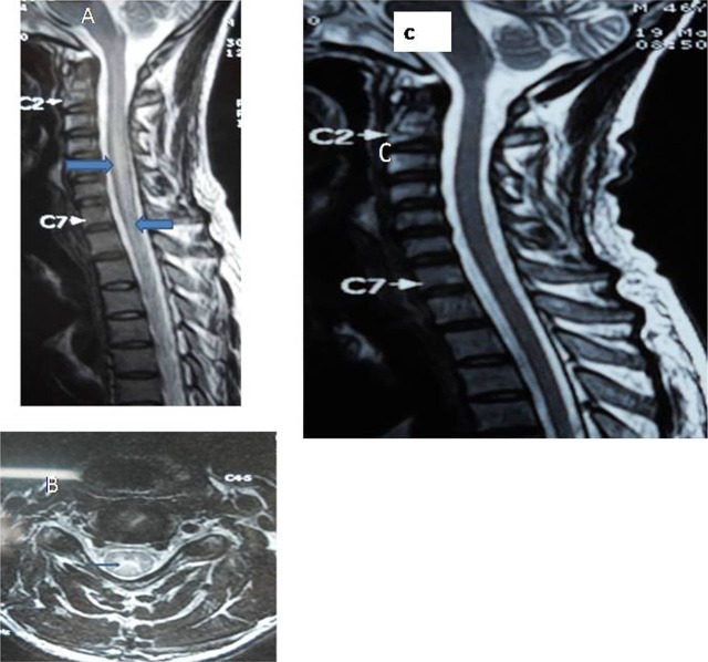
Figure 1.
(A) MRI, cervicothoracic spine T2-weighted sagittal image showed hyperintense signals, extending from cervical first till thoracic second segments with swollen cord. (B) T2-weighted axial image depicted hyperintensities at cervical area. (C) Repeat T2-weighted sagittal image showed complete resolution of the lesion.

