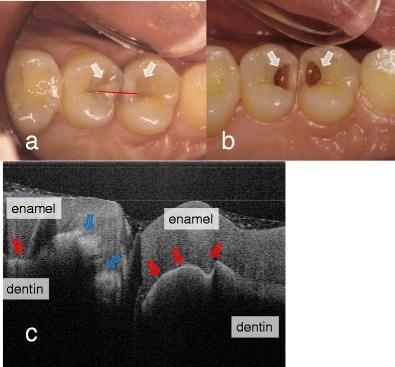
Fig. 1.

Dental caries in first and second premolars. a Occlusal view before the surgical treatment. Underlying dark shadows were visually observed at the first and second premolars (arrow). SS-OCT observation was performed along red line. b Occlusal view during the cavity preparation. Presence of deep lesions with softened dentin was obvious (white arrow). c SS-OCT image at red line in (a) before cavity preparation. Bright zone indicates the increased light scattering in porous demineralized tissue (blue arrow). A strong reflection penetrating along the DEJ indicates the lesion is “cavitated” (red arrow)
