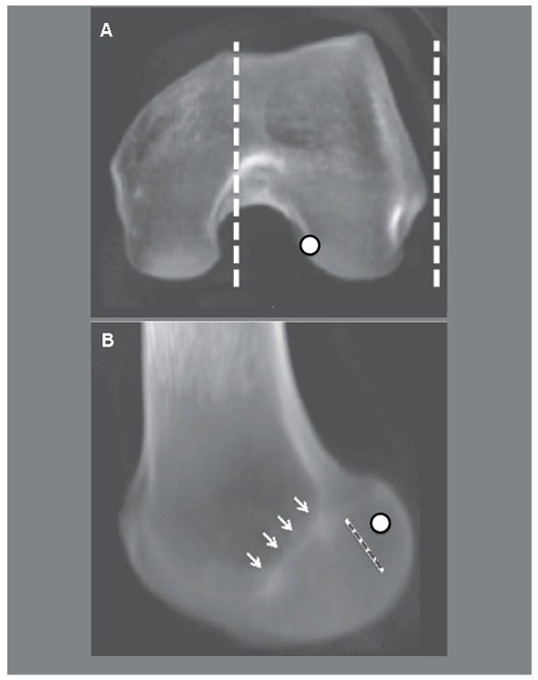
Figure 1. Transparent CT scan of the lateral femoral condyle. A) Dotted line: limit of the lateral femoral condyle including the intercondylar notch. B) Sagittal view of the selected lateral condyle. White arrows: Blumensaat line; dotted line: resident ridge; circle: central ACL footprint.

