
Figure 3.
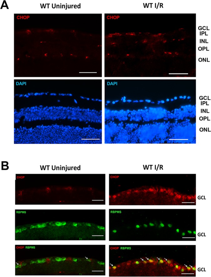
Ischemia/reperfusion injury–induced CHOP upregulation in RGCs. (A) Representative images show CHOP and DAPI staining in WT uninjured and I/R-injured retinas. IPL, inner plexiform layer; OPL, outer plexiform layer. CHOP expression was detected mainly in the GCL, INL, and ONL. In the I/R-injured eye, the CHOP expression appeared to be increased. Scale bar: 100 μm. (B) Representative images of the GCL showing CHOP and RBPMS and DAPI staining in WT uninjured and I/R-injured eyes. Scale bar: 50 μm. Retinal I/R injury increased CHOP expression (red), which was colocalized with RBPMS (a marker of RGC, green) immunoreactivity. Retinal cross-sections were obtained from mouse retinas 3 days after I/R (n = 4).