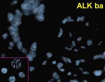
Fig. 2.

FISH of normal breast terminal duct lobule showing normal disomy for ALK [3′-spectrum orange, 5′-spectrum green] signals (yellow); a normal pattern in non-tumor breast cells. Inset CEP2 probe [spectrum aqua] performed on metaphase and control cells with normal 2 signal pattern
