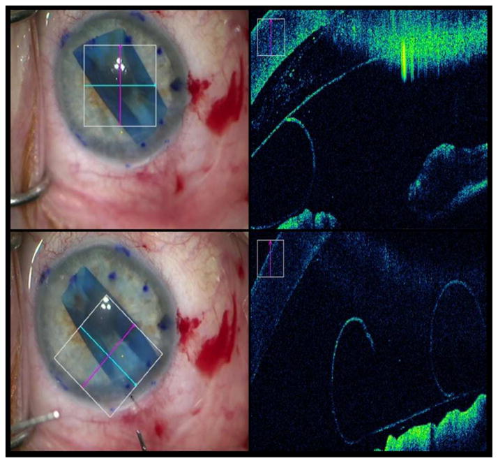
Figure 3. Intraoperative Optical Coherence Tomography Depictions of Graft Orientation During Descemet Membrane Endothelial Keratoplasty.
The left column shows en face views. In the right column, the accompanying intraoperative optical coherence tomography (OCT) images from the en face views. Top row: Inverted graft after insertion that required manipulations to flip it over. Bottom row: Graft orientation corrected after manipulations and verified on intraoperative OCT.

