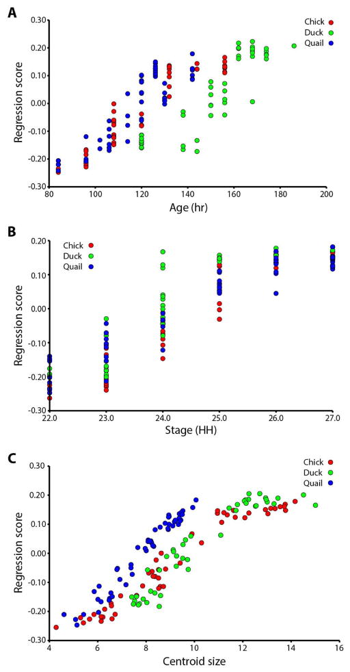
Figure 2. Regression analyses.
A. Regression on age (hours). Duck embryos are separated to the right of the other two species’ age trajectories. B. Regression on stage (HH). Duck embryos have higher shape scores at HH24 and later, compared to chick and quail. C. Regression on centroid size. Quail embryos separate to the left of the other two species’ shape-size curves.

