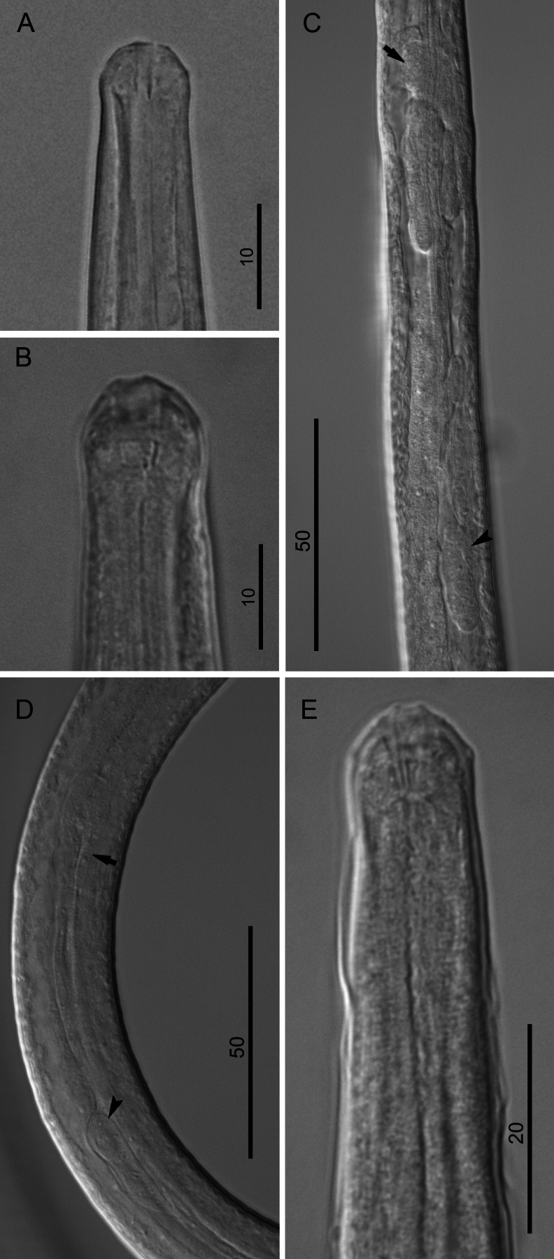
Fig. 2.
Morphology of third-stage larvae of B. malayi. a Cephalic extremity of 3 dpi larva, lateral view. b Cephalic extremity of 5 dpi larva, lateral view; note hyaline tissue around buccal cavity. c Anlage of reproductive system of 5 dpi female larva, right lateral view; note nerve ring (arrow) and genital anlage (arrowhead). d Anlage of reproductive system of 5 dpi male larva, lateral view; note esophago-intestinal junction (arrow) and genital anlage (arrowhead). e Anterior body end 7 dpi male larva, lateral view; note detached third-stage cuticle. Scale bars in micrometers

