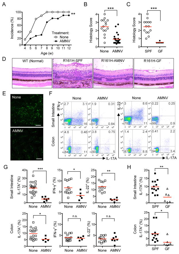
Figure 2. Elimination of commensal microbiota in R161H mice attenuates spontaneous uveitis and reduces the Th17 cells in the gut.
(A) Delayed onset of spontaneous uveitis in AMNV-treated vs. untreated R161H mice by weekly fundoscopic analysis. **p< 0.005 by 2-way ANOVA.
(B) Histology scores of untreated (None) and AMNV-treated R161H mice (9–12 wks old).
(C) Histology scores of age-matched untreated SPF and germ-free (GF) R161H mice (combined data from 7 and 11 wk-old). ***p<0.0001 by Mann-Whitney U test.
(D) Representative histopathology of SPF, AMNV-treated and GF R161H mice (original magnification, 200x).
(E) Expression of GFP in fresh ileum samples of R161H Nr4a1GFP mice treated or not with AMNV. Images are representative single plane of the Z stack. Scale represents 25 μm.
(F) Representative plots of cytokine-producing LP CD4+ T cells from R161H mice treated, or not, with AMNV.
(G) Compiled data for (F) from at least 5 experiments (4–16 wks old, 2–3 mice were pooled per group in each plot). ***p<0.0001, **p<0.005, *p<0.05, n.s., not significant.
(H) Frequencies of IL-17A-producing LP CD4+ T cells from SPF R161H and GF R161H mice. Data compiled from 2 experiments. **p<0.01, *p<0.05. Also see Figures S2 and S3.

