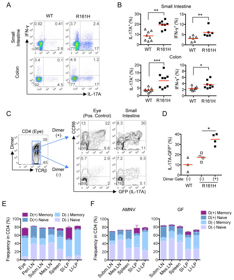
Figure 3. Retina-specific T cells preferentially exhibit an antigen-experienced, Th17 phenotype in the intestine of R161H mice.
(A) The frequency of Th17 cells is enhanced in the intestine of R161H mice compared to their WT littermates. Representative plots of small intestine and colonic LP CD4+ cells from 8-wk old mice for intracellular IL-17A and IFN- γ staining following 4 h ex vivo stimulation with PMA and ionomycin in the presence of Brefeldin A.
(B) Compiled data from several experiments for small intestine and colon (2 mice from each genotype were pooled as a sample). *p<0.05, **p<0.01, ***p<0.001.
(C) IL-17A (GFP) and CCR6 expression of freshly isolated cells from intestinal LP and uveitic eyes in CD4+ p161 Dimer (+) and Dimer (−) populations. One representative experiment of two (in which 2 mice pooled per sample) is shown.
(D) Compiled data from individual small intestines of 3 additional experiments. *p<0.05 by Mann-Whitney U test. Red horizontal lines are group means.
(E, F) Proportions of Memory (CD44hiCD62Llo) and Naïve (CD44loCD62Lhi) within the Dimer (+) or Dimer (−) populations of CD4+ T cells from LP compared to peripheral lymphoid tissues: (E) SPF R161H mice. (F) AMNV-treated and GF R161H mice (CD44loCD62Llo and CD44hiCD62Lhi populations are not displayed). Mean +/− SEM of 4–12 mice is shown.

