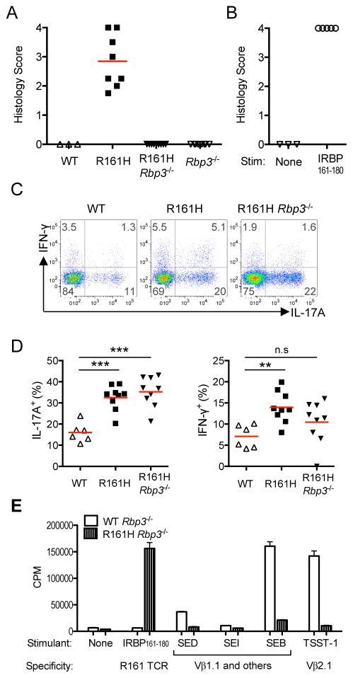
Figure 4. R161H T cell activation in the intestine is not dependent on endogenous IRBP or bacterial superantigen.
(A) Rbp3−/− R161H mice do not develop spontaneous uveitis, due to lack of the target retinal Ag.
(B) Activated R161H Rbp3−/− T cells induce uveitis in WT naïve recipients.
(C) The high proportion of IL-17A-producing CD4+ T cells in the LP compared to WT is maintained in R161H Rbp3−/− mice. Data shown is one of four experiments with similar results.
(D) Compiled data from individual small intestines in 4 additional experiments. **p<0.005, ***p<0.001 by Mann-Whitney U test. Red lines indicate the mean of the group. (E) IRBP-specific T cells do not proliferate in response to commercially available purified enterotoxins with known Vβ specificities. CD4+ T cells isolated from lymph nodes of WT or R161H mice on the Rbp3−/− background were stimulated with 100 ng/ml of the indicated superantigen. SEB: Staphylococcal Enterotoxin B; SEI: Staphylococcal Enterotoxin I; SED: Staphylococcal Enterotoxin D; TSST-1: Toxic Shock Syndrome Toxin-1. Positive control: IRBP161–180 (1 μg/ml). Proliferation was assayed as 3H-thymidine uptake. Also see Figure S4.

