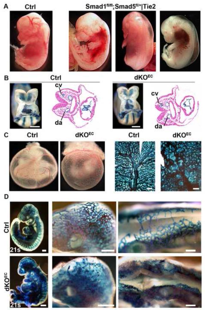
Figure 1. Normal vasculogenesis but impaired angiogenesis in dKOEC embryos.
a, E13.5 control embryo (left panel) and embryos containing one functional allele of Smad1 or Smad5 in ECs. b-d, Embryos lacking the four alleles of Smad1 and Smad5 in endothelium. b, Control and mutant E8.5 whole-mount and sectioned R26R reporter embryo stained with X-gal (blue). c-d, Whole mount and flat mounted X-gal stained yolk sacs of control and mutant E9.5 R26R reporter embryos. In d, magnified views are shown of the head and roof of the hindbrain (middle and right panels). Abbreviations: cv, cardinal vein; da, dorsal aorta; s, somites. Scale bars: 250 μm (b,c,d left panels) and 300 μm (d middle and left panels). See also Figure S1.

