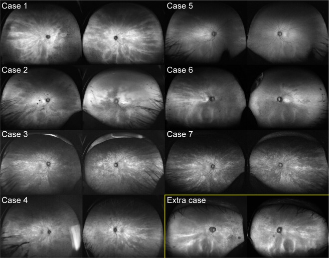
Figure 2.
Peripheral radial FAF.
Notes: Except in case 5, the radial FAF is depicted better in the periphery than at the posterior pole. The images within the yellow box represent those of the study participant with a radial pattern limited to the periphery.
Abbreviation: FAF, fundus autofluorescence.

