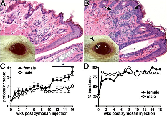
Fig. 2.

Evaluation of blepharoconjunctivitis development in SKG mice. a Photograph and stained histological section of a naive, healthy control BALB/c mouse. b Photograph and stained histological section of an SKG mouse that had received a zymosan injection, showing eyelid inflammation (blepharitis) and conjunctival discharge. Arrowheads indicate thickened lid margin, which coincides with epithelial hyperplasia and leukocytic infiltration visualized by histopathology (arrows). The presence of such periocular manifestations in SKG mice was graded over time (0–16 weeks after zymosan injection) using a binary scoring system to assay for the complete absence (score of 0 as shown in panel a), the presence of blepharitis (score of 1) or blepharitis with coinciding conjunctivitis and/or discharge (as shown in panel b; score of 2). Total score per mouse (sum of both eyes) (c) and percentage incidence (score ≥1; panel d) were plotted as a function of time since zymosan injection. A prominent response (p < 0.05) was observed for both male and female mice that received a zymosan injection versus SKG mice that did not receive a zyomsan injection (c). Ψ p < 0.05 indicates effect of sex among SKG mice that received a zymosan injection; n = 12–18 mice/group (combined two individual experiments). BALB/c mice were included as a negative control group (scores = 0)
