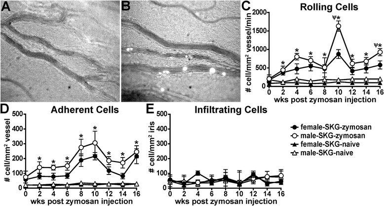
Fig. 3.

Intravital videomicroscopy reveals cellular rolling and adhesion, but not infiltration, within the microvasculature of the eye. Photographs show representative still images from videomicroscopy of the iris vasculature and rhodamine-labeled hematopoietic cells from uninflamed eyes of naive SKG mice (a) and from inflamed eyes of SKG mice that had received a zymosan injection (b). The iris vasculature and tissue were imaged biweekly over a 16-week period in naive SKG mice or in SKG mice that had received a zymosan injection, and videos were analyzed offline for numbers of rolling, adherent and infiltrated cells per square millimeter of vessel or iris tissue (c–e). *p < 0.05 indicates comparison between zymosan and naive SKG mice within each sex; Ψ indicates sex effect within SKG mice that had received a zymosan injection; n = 12–18 mice/group (combined two individual experiments)
