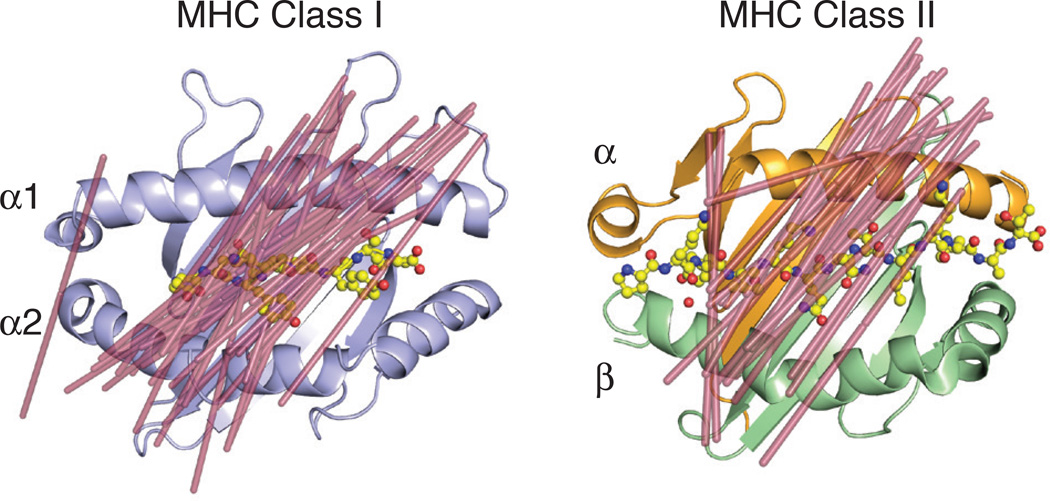
Fig. 2. MHC class I and class II restricted TCR docking orientations.
Shown are the docking orientations of αβ TCRs on their MHC class I (left, PDB ID: 2CKB) or class II (right, PDB ID: 1FYT) ligands. Lines were drawn (shown in raspberry) from the two external conserved cysteines in the Variable Ig domains (C22 of α chain and C23 of β chain) to demonstrate orientation of the TCR on the MHC surface. The following PDB IDs were used for the complex structures for the class I model: 1FO0, 1KJ2, 3RGV, 2CKB, 2OI9, 3PQY, 2OL3, 1AO7, 1BD2, 1LP9, 1OGA, 2BNQ, 3GSN, 3HG1, 3O4L, 3QDJ, 3QDM, 3UTS, 3VXM, 3VXR, 3VXU, 4G8G, 2NX5, 3MV7, 2AK4, 4JRY, 3DXA, 3KPS, 2YPL, 1MI5, 3FFC, 3SJV, 4MJI, 2ESV; and for the class II model: 3PL6, 4OZF, 4OZI, 4GG6, 4E41, 2IAN, 1FYT, 4H1L, 2WBJ, 1YMM, 1ZGL, 3O6F, 4P4K, 3C5Z, 3C60, 3C6L, 3RDT, 3MBE, 2PXY, 1U3H, 3QIB, 1D9K.

