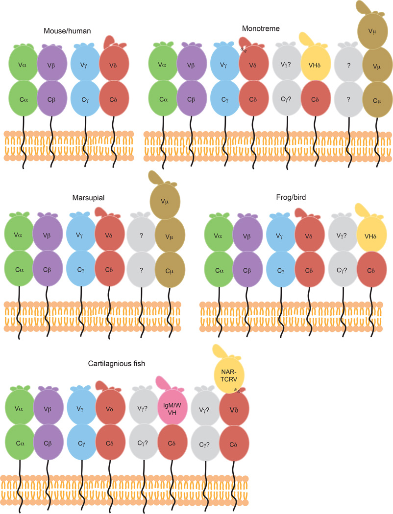
Fig. 8. Comparison of TCR chains present in diverse species.
Cartoon diagrams of the variable TCR chains present in a variety of species including mice and humans, monotremes, marsupials, frogs and birds, and cartilaginous fish. Traditional TCRα (green), β (purple), γ (blue), and δ (red) chains are found in all species. TCRδ chains with VH-like TCRVδ domains are shown in yellow, and those expressing true IgVH domains via transrearrangements in pink. Potential binding partners for unusual TCRδ chains are colored grey. Predicted structure of three-domain TCR chains, NAR- TCR and TCRµ (brown) are shown in B, C, and E, with the NAR-TCRV domain depicted yellow due to its similarity with IgNARV domains. CDR1, 2, and 3 loops depicted at the top of each TCRV domain, with larger loops representing longer CDR3s found in some Vδ domains. Predicted disulfide linkages due to extra cysteine residues in monotreme TCRδ CDR3 (B) and between terminal and supporting shark NARTCRV domains (E) are labeled.

