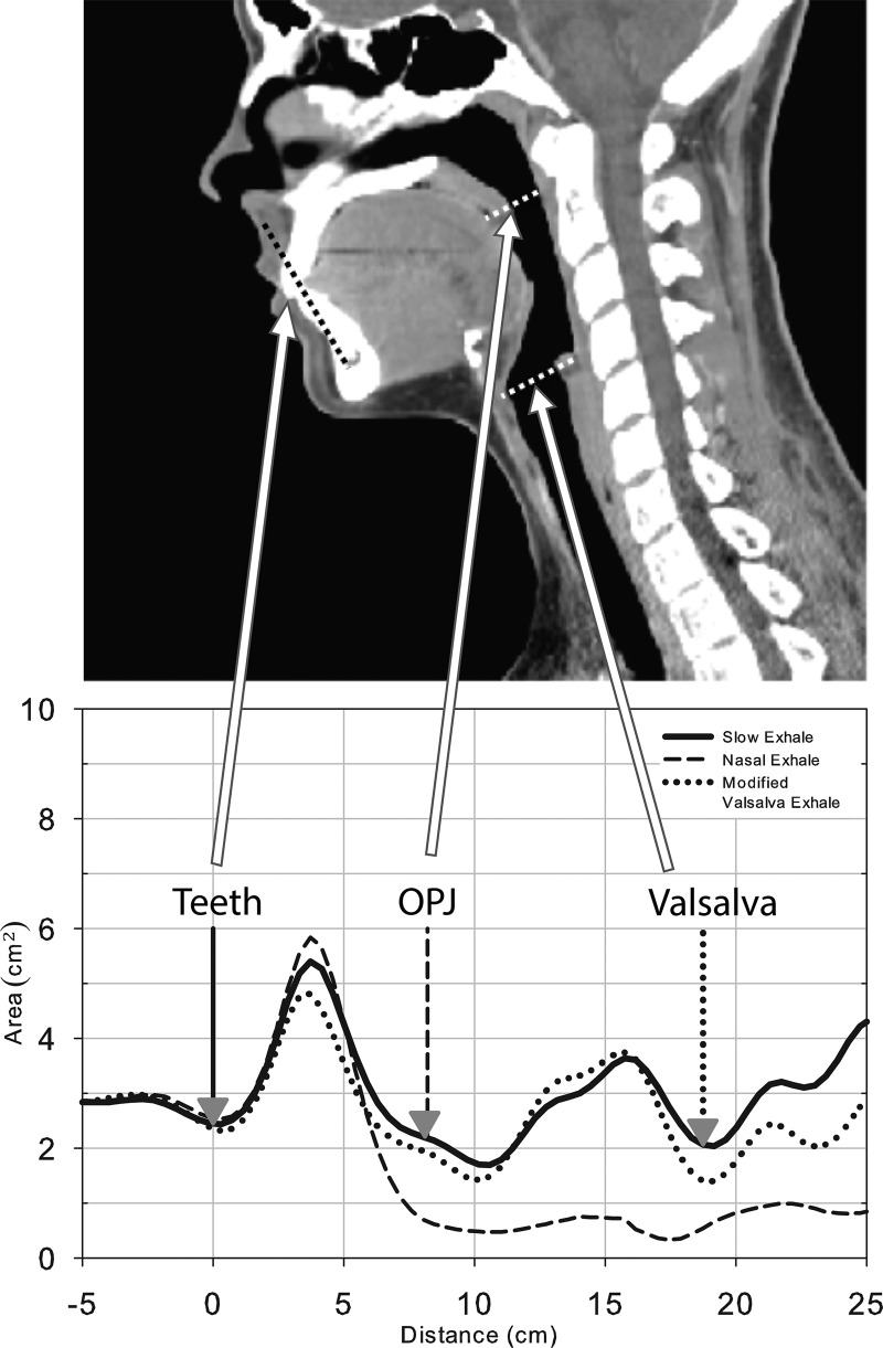
FIG. 2.
A visual representation of the anatomic measures on a pharyngogram (bottom) with corresponding anatomic landmarks on a midsagittal CT scan (top). The CT is for illustrative purposes and representative of normal breathing, not any of the APh breathing conditions. The bottom display is a schematic of three pharyngograms displaying the three different breathing conditions superimposed to illustrate how the nasal and modified valsalva pharyngograms are, respectively, used to locate the OPJ and the glottis on the slow exhale pharyngogram.

