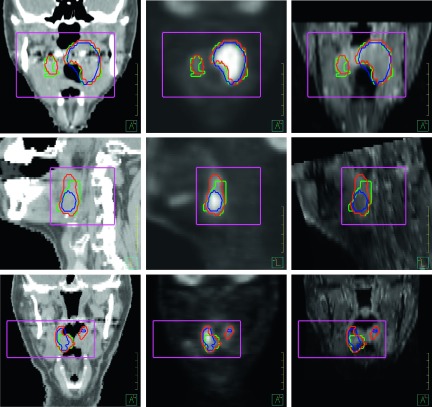
FIG. 5.
The multichannel segmented contours (red) and the PET-based segmented contours (blue) were compared with the physician-defined manual contours (green). The rectangular boxes manually drawn in the preprocessing step define a small region for the segmentation. Left to right: contours overlaid on CT, PET, and MR images. Top to bottom: coronal, sagittal, and coronal views of the target volumes for patients 3, 16, and 1, respectively.

