Figure 3.

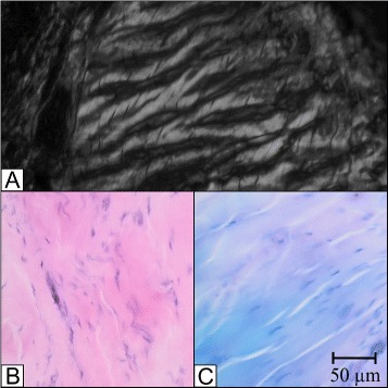
Histology of the ALL. Different histological illustrations of the ALL; A: Polarization with crimping; B: HE stain; C: Giemsa stain.

Histology of the ALL. Different histological illustrations of the ALL; A: Polarization with crimping; B: HE stain; C: Giemsa stain.