
Figure 2.
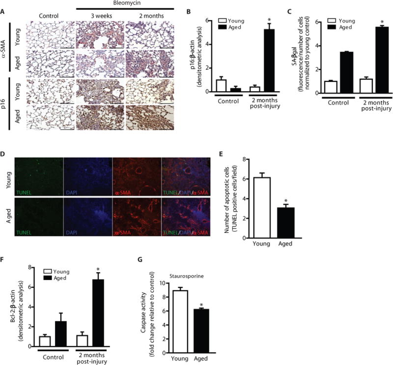
Impaired fibrosis resolution in aged mice is associated with myofibroblast senescence and apoptosis resistance. Young and aged mice were subjected to bleomycin-induced lung fibrosis. Lung tissue was harvested or cells were isolated at various time points following injury. (A) Immunohistochemical analysis of α-SMA expression; myofibroblast marker (upper panels), and p16 expression; senescence marker (lower panels). (B,F) Whole lung tissues were analyzed at the time points indicated by Western blot for protein expression and densitometric analyses of p16 (B) and Bcl-2 (F) was performed; *p value < 0.05, compared with all other groups. (C) Fibroblasts from young and aged mice (uninjured and 2 m post-injury) were isolated and cultured ex vivo. Senescence was evaluated by quantitative measurement of senescence-associated β-galactosidase (SA-βgal) activity; *p value < 0.05, compared with all other groups. (D–E) Lung tissue harvested at 3 weeks post-injury was assessed by immunofluorescence for TUNEL and α-SMA expression (D), and apoptotic cells were quantified by counting the number of TUNEL-positive cells/field in >50 random fields of view (E); *p value < 0.05, compared with young. (G) Fibroblasts were isolated from young and aged mice 6 weeks post-injury and cultured ex vivo. Cells were treated with or without staurosporine (300 nM) for 5 h and caspase activity was assessed. Values represent mean ± s.e.m.; n = 3–5; *p value < 0.05, compared with young. Scale bars, 100 μm.