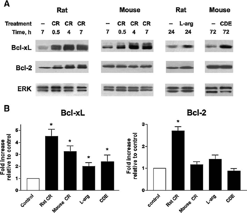
Fig. 1.
Pancreatic levels of prosurvival proteins Bcl-xL and Bcl-2 are up-regulatedin rodent modelsof acute pancreatitis. Pancreatitis was induced in rats and mice as described in Materials and methods by administration of cerulein (CR), L-arginine (L-arg) or choline-deficient ethionine supplemented (CDE) diet. Control animals received saline injections (in the CR and L-arg models) or control diet (in the CDE model). (A) Bcl-xL and Bcl-2 protein levels were measured by Western blot analysis in pancreatic tissue of control and pancreatitic animals at indicated times after the induction of pancreatitis. Blots were re-probed for ERK1/2 to confirm equal protein loading. In this and other figures, Western blot data represent experiments that were repeated with similar results on at least three animals in each group. (B) Western blot was performed as in (A) on pancreatic tissue from rats and mice with fully developed pancreatitis: at 7hintheCR models,24 h in the L-arg model, and 72 h in the CDE model. The intensities of Bcl-xL and Bcl-2 bandson the immunoblots were quantifiedby densitometry and normalizedtothatofERK1/2 (loading control) inthe same sample. The mean ratio of Bcl-xL/ ERK1/2 (or Bcl-2/ERK1/2) intensities in animals with pancreatitis was further normalized to that in control group at the same time point. Values are means±SE from at least 3 animals per group. *p<0.05 compared with control.

