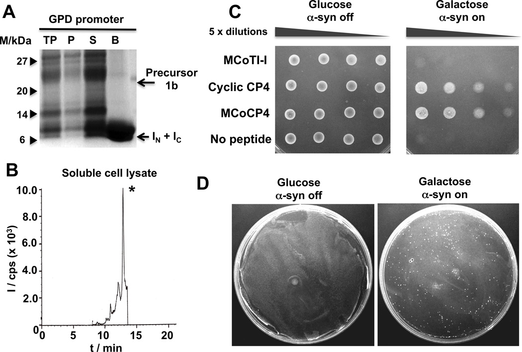
Figure 3.
Expression of cyclotide MCoCP4 in S. cerevisiae cells using Npu DnaE intein-mediated PTS. A. SDS-PAGE analysis of the recombinant expression of cyclotide precursor 1b in S. cerevisiae strain W303-1a cells for in-cell production of cyclotide MCoCP4 (lane description as in Fig. 2). B. Analytical HPLC-MS/MS trace of the soluble cell extract fraction from S. cerevisiae cells expressing precursor 1b under the control of a GPD constitutive promoter. The peak marked with an asterisk corresponds to folded cyclotide MCoCP4. MS/MS analysis was performed using the ion (m/z = 1038.8) for both Q1 and Q3 (units are as described in Fig. 2B). C. Spotting assays of normalized, serially diluted cells of the synucleopathy model expressing cyclotides MCoTI-I (negative control) and MCoCP4, and cyclic peptide CP4 (positive control). D. Phenotypic screening using a yeast synucleopathy model. Screening cells were transformed with a mixture of p426GPD plasmids encoding cyclotides MCoCP4 (cytoprotective) and MCoTI-I (inactive) in a ratio of 1 to 5×10⁴. Glucose and galactose plates are shown to illustrate how a single bioactive cyclotide can be isolated from more than 107 transformants on a single plate.

