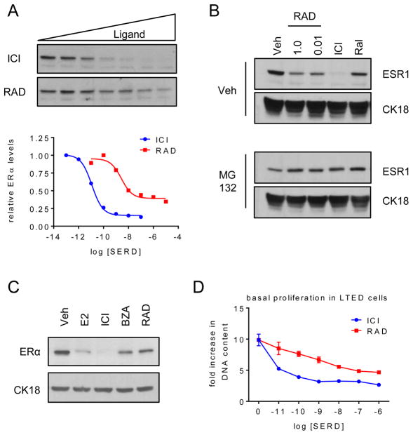
Fig. 2. RAD1901 downregulates ESR1 expression through receptor degradation.
A) MCF7 cells were treated for 24 hours with ICI (10−13–10−7 M) or RAD (10−11–10−5 M). Expression of ESR1 and loading control cytokeratin 18 (CK18 – Supplementary figure 1A) in whole cell extracts were detected by immunoblot (top). ESR1 levels relative to CK18 were quantitated by densitometry using Adobe Photoshop (bottom). B) MCF7 cells were plated as in Fig. 1B prior to 1 hour pre-treatment with vehicle or MG132 (10 μg/ml), followed by 6 hours of treatment with 10−7 M vehicle, ICI, Ral or RAD (10−8 or 10−6 M). ESR1 expression was detected as in (A). C–D) LTED MCF7 cells were plated in phenol red free media supplemented with FBS that was stripped of growth factors twice using charcoal. C) After 48 hours, cells were treated for 24 hours with E2 (10−7 M) or SERDs (10−6 M) and ESR1 was analyzed as in (A). D) LTED MCF7 cells were treated with ICI or RAD (10−11 – 10−6 M) on days 1, 4, and 6 of an 8 day proliferation assay and analyzed as in Fig. 1.

