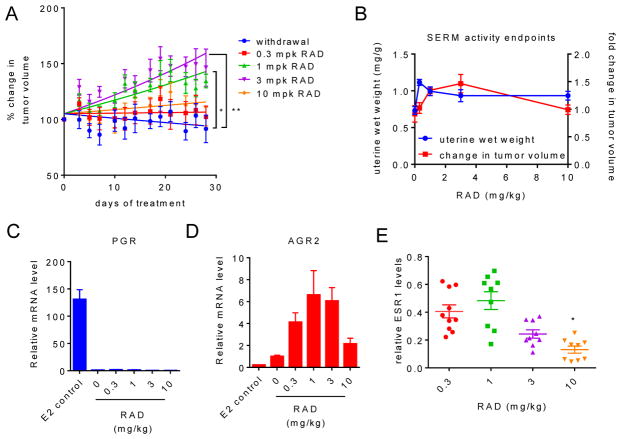
Fig. 4. RAD1901 exhibits dose dependent growth stimulation of MCF7 xenograft tumors.
MCF7 xenograft tumors were initiated in ovariectomized female nu/nu mice as in Fig. 1. Estrogen pellets were surgically removed when tumors reached ~0.1 cm3 volume, and animals (n = 6–10) then received daily treatment with vehicle or RAD (0.3–10 mg/kg sc). Mean tumor volume +/− SEM per day of treatment is presented. Significance as compared to the vehicle (2-way ANOVA of matched values followed by Bonferroni comparison) is indicated (* p < 0.05, ** p < 0.0005). B) Uterine wet weight at sacrifice (measured as in Fig. 3) and % change in tumor volume (as compared to size at randomization) calculated using the final measurement recorded for mice in (A) are graphically presented. C–D) Expression of ESR1 target genes in tumors was analyzed essentially as in Fig. 1. Estrogen only samples from Fig. 1D were included for comparison. E) ESR1 levels in tumor tissues were analyzed as in Fig. 3 and were normalized to similarly detected Lamin-A. Significant downregulation (* p < 0.05) of ESR1 was determined by ANOVA followed by Bonferroni comparion.

