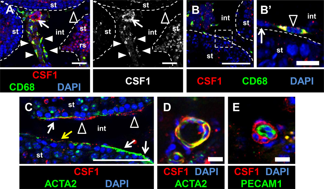
Figure 5. CSF1 protein is localized to macrophages and perivascular smooth muscle cells in the adult testis.
CSF1 protein is detected within interstitial macrophages (A, white arrowheads), peritubular macrophages (A-C, black arrowheads), and vascular-associated cells (A, arrow), but is absent or weakly expressed in ACTA2-positive peritubular myoid cells (white arrows in B’, C; yellow arrow in C points to myoid cell with weak CSF1 expression). CSF1 is detected in round spermatids (“rs” in A). Panel to the right of A is the CSF1-only channel from the image in A. Labeling perivascular smooth muscle cells with anti-ACTA2 antibody (D) or endothelial cells with anti-PECAM1 (E) reveals that vascular-associated CSF1 is specifically localized to perivascular smooth muscle cells. All images are from cryosectioned testes. Thin scale bar, 50 µm; thick scale bar, 10 µm.

