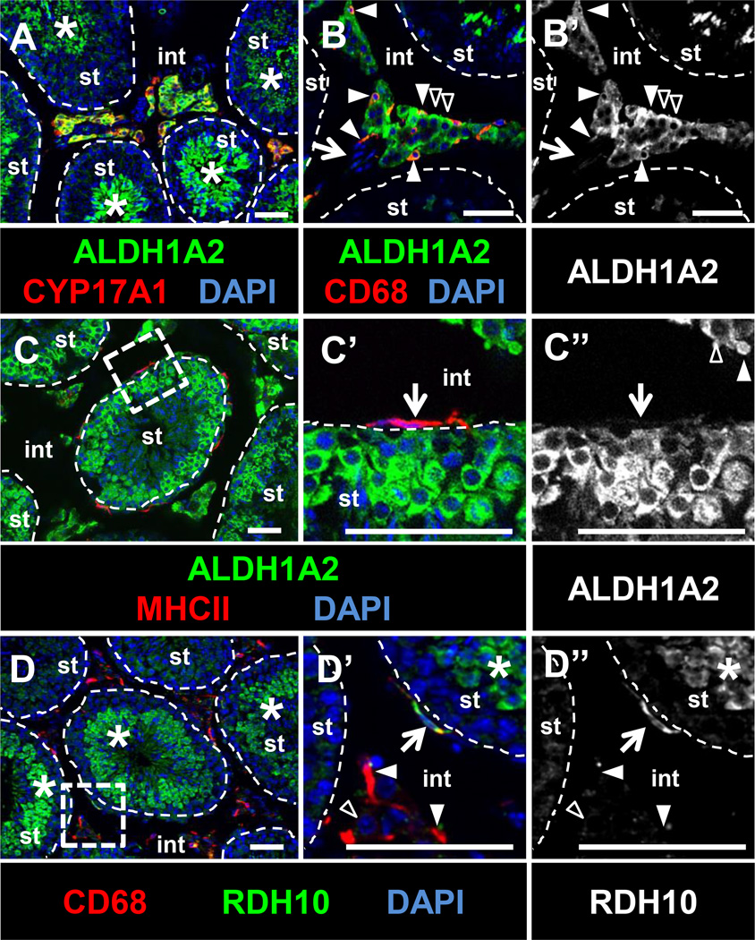
Figure 6. RA synthesis enzymes ALDH1A2 and RDH10 are expressed in testicular macrophages.
(A–C) ALDH1A2 is detected within CYP17A1-positive Leydig cells (black arrowheads in B), interstitial macrophages (CD68-positive; B, white arrowheads), and germ cells (asterisks in A). ALDH1A2 is not expressed in vasculature (B, arrow), and is weakly expressed in MHCII-positive peritubular macrophages (C’, arrow). RDH10 is detected in peritubular macrophages (D’, arrow), but not in most interstitial macrophages (white arrowheads in D’) or Leydig cells (black arrowhead in D’). RDH10 is detected in spermatids and other germ cells (D, D’, asterisks). C’ and D’ are higher magnifications of the boxed regions in C and D, respectively. B’ and C” are ALDH1A2-only channels for B and C’, respectively; D” is the RDH10-only channel for D’. All images are from cryosectioned testes. Scale bar, 50 µm.

