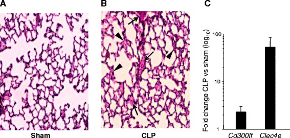
Fig. 2.

Histopathology of CLP affected lungs and expression pattern of novel ARDS candidate genes. Panels a-b : Light micrographs of H&E stained lung sections from sham (a) and CLP (b) mice. The lung histopathology in CLP-challenged mice demonstrates broadening of alveolar septa with sparse monocyte infiltration (arrowheads) and hemorrhage in septa (arrow). Original magnification, 200×. Panel c : The expression of Clec4e and Cd300lf genes in whole mouse lung is represented by horizontal bars. The error bars are standard deviations among three samples. The real time PCR was conducted using commercially available TaqManTM reactions
