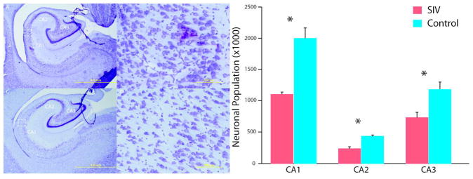
Figure 1.

Hippocampal neuronal loss. SIV infected infants have apparent enlarged ventricles and thinned pyramidal neuronal layers (A, B) compared to control subjects (C, D). In these cresyl violet stained sections, neurons can be differentiated from glia based on a clearly visible nucleolus surrounded by cytoplasm. The CA fields were delineated on the basis of cyto- and chemoarchitecture, and equidistant sections were evaluated throughout the entire length of the hippocampus. Design-based stereology of the hippocampal CA subregions found an overall 42% neuronal reduction. There were no overall volume differences in hippocampus. Magnifications of (A, C) 1.25× and (B, D) 20×; scale bars = 5 mm and 200 μm, respectively. Figure adapted from Curtis et al., 2014.157
