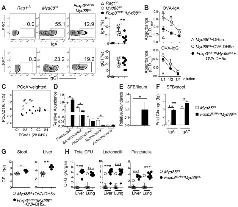
Figure 5. TR-cell specific MyD88 deficiency promotes dysbiosis and and bacterial translocation to deep tissues.
A. Flow cytometric analysis and frequencies of IgA- and IgG1-coated bacteria in the fecal pellets of Rag1−/−, Foxp3EGFPcreMyd88Δ/Δ and MyD88fl/fl mice. B. OVA-specific luminal IgA and IgG1 concentrations in mice (panel A) gavaged with OVA-DH5α bacteria. C. Weighted Unifrac plot of ileal mucosa-associated microbial communities, visualized with principal coordinate analysis (PCoA), in Foxp3EGFPcreMyd88Δ/Δ versus Myd88fl/fl mice (p-value = 0.001 by AMOVA). D. Relative abundance of different microbial phyla in ileal mucosa-associated microbiota in Foxp3EGFPcreMyd88Δ/Δ and Myd88fl/fl mice (*false discovery rate<0.05). E. Relative abundance of SFB taxa in illeal mucosa-associated microbiota in Foxp3EGFPcreMyd88Δ/Δ and Myd88fl/fl mice. F. Real time PCR analysis of SFB 16S rRNA in IgA+ and IgA− fecal bacterial fractions of Myd88fl/fl and Foxp3EGFPcreMyd88Δ/Δ mice. G. Stool (left panel) and liver (right panel) OVA-DH5α bacterial load [expressed as log colony forming units (lg CFU)/g] in Foxp3EGFPcreMyd88Δ/Δ and Myd88fl/fl mice treated with the OVA-DH5α bacteria by oral gavage. H. Loads of total bacteria, Lactobacilli and Pasteurella (all expressed as CFU/g tissue) in the lung and liver of Myd88fl/fl and Foxp3EGFPcreMyd88Δ/Δ mice. Results are representative of 3 independent experiments. Results representative of 3 independent experiments. N=5 mice/group. *p<0.05; **p<0.01; by Student’s unpaired two tailed t test.

