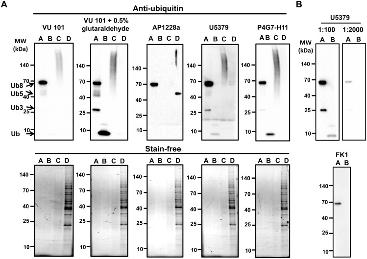
Fig 2. Validation of anti-ubiquitin antibodies.
VU101 in the presence and absence of 0.5% glutaraldehyde pre-treatment, U5379, AP1228a, or P4G7-H11 were used to detect ubiquitin and ubiquitinated proteins. A) Western blot of polyubiquitin chains (Ub3, Ub5, Ub8) (lane A), purified ubiquitin (lane B), polyubiquitinated proteins from H9c2 cells treated with 10μM MG-132 for 36 h obtained from affinity purification using TUBEs (lane C), and unbound fraction from H9c2 cells after removal of polyubiquitinated proteins (lane D). B) Upper figure, Western blot of free ubiquitin (lane A) and polyubiquitin chains (lane B) with U5379 antibody diluted at 1:100 and 1:2000. Lower figure, Western blot of free ubiquitin (lane A) and polyubiquitin chains (lane B) with FK1 antibody diluted at 1:1000 in BSA. Even when the blots were imaged for long time periods no additional bands were seen.

