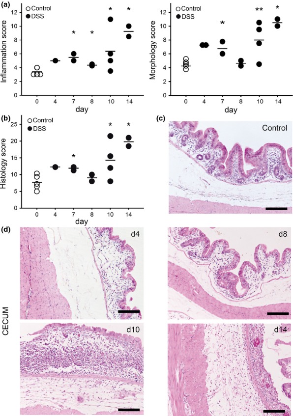
Figure 3.

Histopathological changes of the caecum at different time points after colitis induction. HE-stained caecum sections were scored (a, 1–4) for markers of inflammation (infiltration of lamina propria eosinophils, lamina propria lymphocytes, intraepithelial lymphocytes) and for the distortion of morphological features (villous stunting, villous epithelial injury, crypt distortion). Single parameters were summarized to a global score (b). Black dots represent DSS-treated rabbits (●); white dots represent control rabbits (○). Horizontal lines represent the arithmetical mean; Mann–Whitney test, **P ≤ 0.05, *P ≤ 0.1. Representative HE-stained caecal sections of control rabbits (c) and of DSS-exposed rabbits at different time points after colitis induction (d). Scale: 200 μm.
