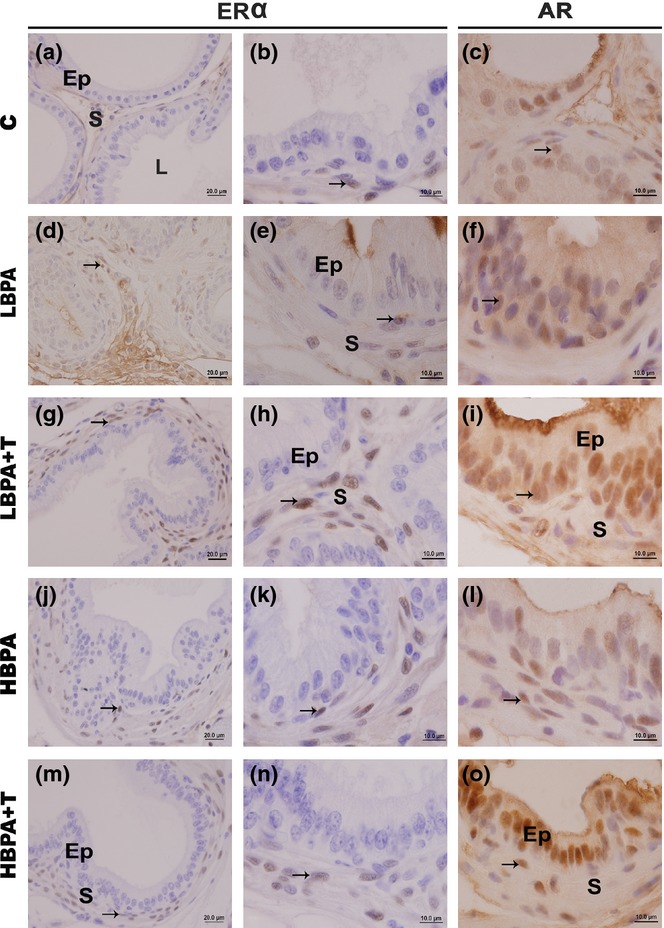
Figure 2.

Immunohistochemistry for oestrogen receptor-alpha (ERα) and androgen receptor (AR) in female gerbil prostates. (a–n) The immunohistochemical analysis for ERα showed a pattern of nuclear staining in the stromal cells (S) of all examined groups (arrows). It is noted, however, that in all treated groups, the immunolabelling was more frequent and evident. (c–o) The immunolabelling for AR was observed in the nucleus of the secretory epithelial cells (Ep) and in the nucleus of fibroblasts and smooth muscle cells in the stroma (S) of all experimental groups (arrows). (Scale bar: 20 μm – Figure 2a, d, g, j, m; scale bar: 10 μm – Figure 2b, e, h, k, n; scale bar: 10 μm – Figure 2c, f, i, l, o).
