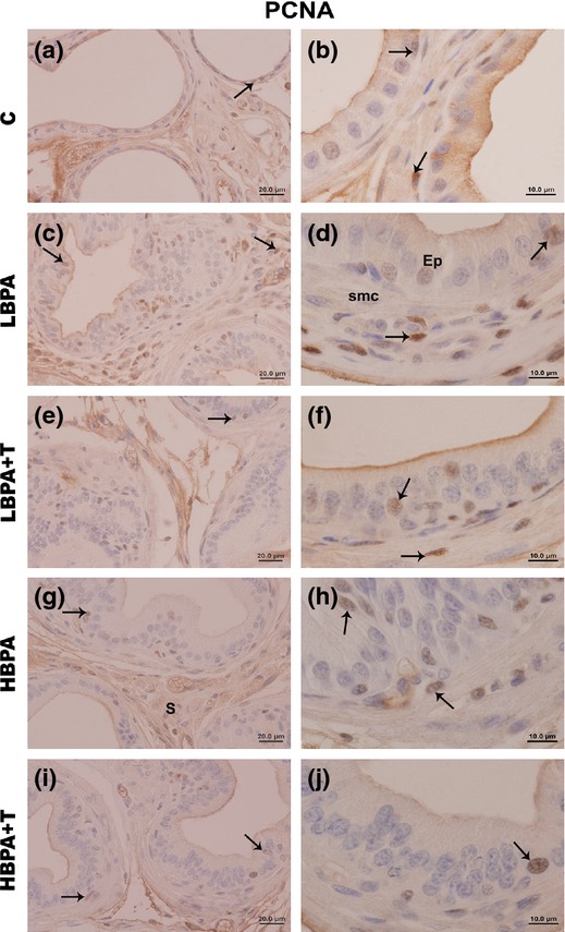
Figure 4.

Immunohistochemistry for proliferation (PCNA) in the female prostate gland of all experimental groups. Immunolabelling for PCNA (arrows) is present in epithelial and stromal cells, particularly in the regions of stratification in the prostate of the LBPA, LBPA + T, HBPA and HBPA + T groups. (Scale bar: 20 μm – Figure 4a, c, e, g, i; scale bar: 10 μm – Figure 4b, d, f, h, j).
