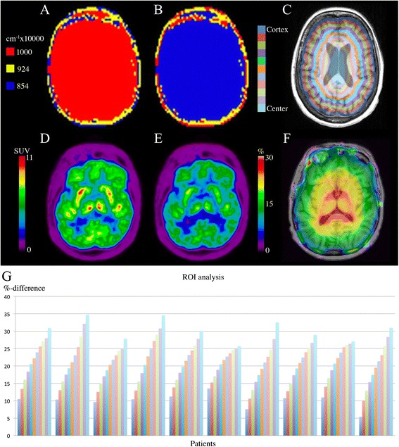
Figure 4.

Effect on PET of simulated fat/water inversion. (A) MR-ACDixon and (B) MR- ACInverted, (C) Annuli drawn on PETDixon fused onto T1w MPRAGE, (D) PETDixon and (E) PETInverted, (F) Δ% (PETDixon, PETInverted) fused onto T1w MPRAGE. (G) ROI analysis of the 11 annuli drawn on the ten FDG brain patients. Color code corresponds to coloring on (C). Notice the visual differences between the PETs in (D) and (E) and the radial gradient in (F) which corresponds to the results in (G). The patient in (A-F) is the third from the left in (G).
