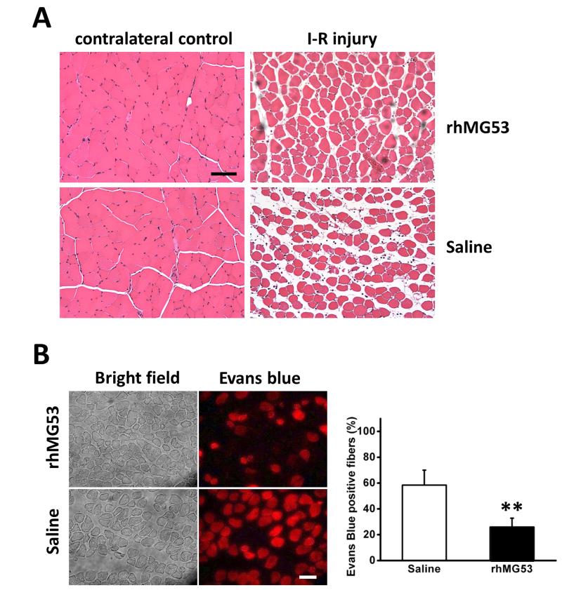
Figure 3. rhMG53 improves muscle structure associated with I-R injury.
(A) H&E staining of control (left) and injured (right) muscles. More necrotic fibers were seen by light pink staining in saline control muscle than that in MG53 treated muscle. n=3 pairs. (B). Fluorescent images of Evans blue positive fibers (right) showed less injury in rhMG53 treated muscle than control. Percentage of total damaged fibers is summarized in the lower panel. The data are mean ± SEM **: P<0.01. n=4 pairs. Scale bars, 50 μm.

