
Figure 2.
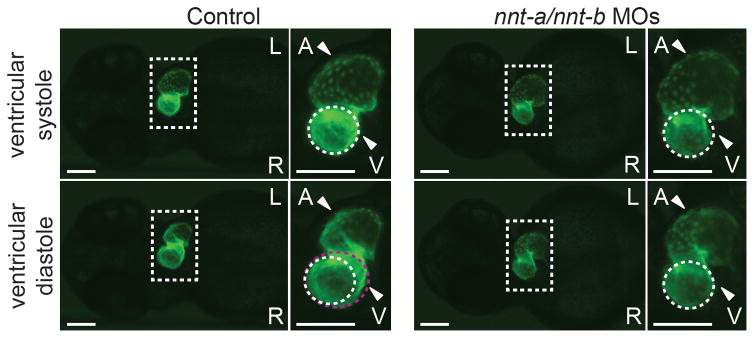
nnt-a/nnt-b morphants display contractile dysfunction. Representative live ventral views of 2 dpf cmlc2:GFP larvae at ventricular systole (top panels) and ventricular diastole (bottom panels). Ventricles in control larvae expand normally as the atrium contracts (compare white dashed circle to violet dashed circle in the bottom inset); ventricles in nnt-a/nnt-b morphants fail to expand during atrial contraction (5 ng each MO injected/embryo). Dashed box corresponds to the magnified image to the right of each large panel; L, left; R, right; A, atrium; V, ventricle; scale bars, 100 μm. nnt-a/nnt-b morphants also display brachycardia (mean 91.5 vs. 66.8 beats/minute, controls vs. morphants; p<0.0001; student’s t-test; n=10 larvae/injection, repeated twice; see Supplemental movies).