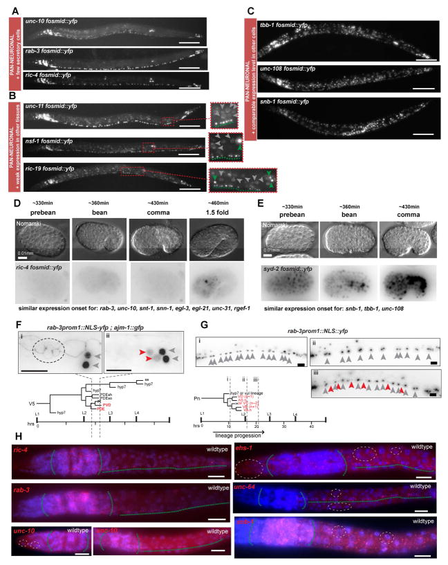
Fig. 2. Different Categories of Pan-neuronal Genes.
Pan-neuronal genes can be grouped in three categories based on their expression in non-neuronal cells (panels A – C).
A: Expression in all neurons and only few non-neuronal secretory cells.
B: Expression in all neurons and weaker expression in other tissues. Expanded boxes show better the difference in levels between neurons and non-neuronal cells. Green arrowheads indicate neurons, dashed greens line underlines ventral nerve cord motorneurons (VNC MNs) and grey arrowheads indicate non-neuronal cells.
C: Expression in all neurons and equally bright expression in all other tissues. Fluorescent images of L4/young adult worms of selected fosmid reporter for each category are shown. For description of spatial expression patterns of all fosmid reporters see Fig. 1C. Temporal onset of expression of pan-neuronal genes differs between genes that belong in different categories (panels D – G).
D: Embryonic expression onset of the fosmid reporter of ric-4, a pan-neuronal gene that is more restricted to the nervous system. Expression at first is detected at the comma stage, when all neurons have already been born. Other pan-neuronal genes that are also mainly nervous system restricted (listed below) have similar temporal expression pattern.
E: Embryonic onset of expression of the fosmid reporter of syd-2, a pan-neuronal gene that is expressed broadly in non-neuronal cell types. Broad expression is detected in very early embryonic stages when neurons are not yet born. Other pan-neuronal genes that are also expressed broadly outside the nervous system (listed below) have similar temporal expression pattern.
F – G: Onset of expression of the neuronal restricted rab-3 in post-embryonically born neurons. In F, the V5 postembryonic lineage gives rise to two neurons, PDE and PVD, two glial cells and epidermal cells. rab-3prom1::2xNLS-yfp expression is detected only in mature postmitotic PDE and PVD neurons (ii), but not at an earlier stage in the “young” postmitotic PDE neuron and the PVD progenitor (i). Also in ii, the YFP expression levels in PDE and PVD (red arrowheads) are lower in comparison to neighboring neurons SDQL and PVM (grey arrowheads) that are born in the embryo. In later larval and adult stages PDE and PVD expression of rab-3prom1 is similar to the expression in SDQL and PVM. ajm-1::gfp is an apical junction marker that is used to follow the different stages of progression of the V5 lineage. In (i) the dashed circle indicates the ajm-1::gfp expression in 4 cells at the corresponding stage (i) indicated in the lineage diagram. One of these 4 cells is the “young” PDE neuron. In G, the Pn postembryonic lineage gives rise to different VNC MN types. Expression of rab-3prom1::2xNLS-yfp is not detected in the neural progenitors (i), or even at a stage when the neurons have just been born (ii). YFP expression in the postembryonic VNC MNs (read arrowheads) is detected only at a later stage (iii) and is initially weaker in comparison to YFP expression of the embryonically born VNC neurons (grey arrowheads). In later larval stages and adult worms all VNC neurons have more similar rab-3prom1::2xNLS-yfp expression levels. In F and G, grey arrows indicate embryonic neurons and red arrows indicate postembryonic neurons.
H: Single molecule in situ hybridization (smFISH) verifies expression patterns of selected pan-neuronal genes. C. elegans larvae were fixed and hybridized at the L1 stage. In red the labeled smFISH probes and in blue is DAPI staining. smFISH for ric-4, rab-3 and unc-10 (left column) shows neuronally restricted fluorescent signals. smFISH for unc-10 recapitulates the unc-10 fosmid reporter expression in just a few cells in the tip of the head (dashed white circle). smFISH for ehs-1, unc-64 and snb-1 shows more broad staining in cells outside the nervous system corroborating the fosmid reporter results. Green dashed lines outline nervous system (head ganglia and VNC). White dashed-line circles outline examples of expression in non-neuronal cells.
Scale bars are 0.1 mm in A – C and 0.01 mm in D – H.

