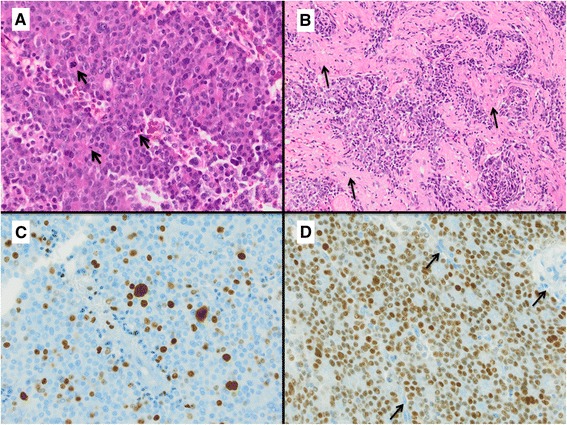
Fig. 1.

Diagnostic criteria for atypical pituitary adenomas. An example of an atypical pituitary adenoma (ACTH-cell adenoma) with several mitotic figures (Arrows in a) in one HPF (HE staining, magnification 400x), infiltration of surrounding meninges (Arrows in b, HE staining, magnification 100x), Ki67 index >4 % (c, magnification 400x) and strong nuclear p53 expression in >3 % of cells (d, magnification 400x; Arrows: negative endothelial cells indicating antibody specificity)
