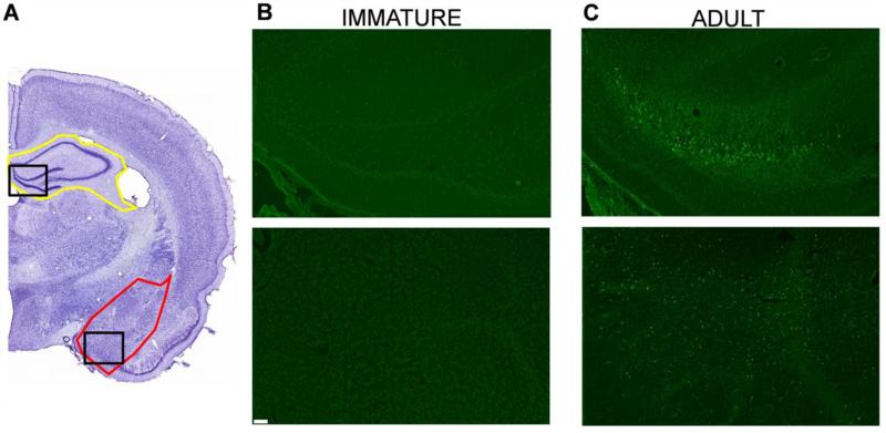
Figure 7. Immature rats do not suffer neuronal degeneration, 1 day and 7 days after soman-induced SE.
A. Cresyl violet photomicrographs outline the brain regions (amygdala in red; hippocampus in yellow) from where the FJC photomicrographs (B and C) were taken (the specific areas shown in the photomicrographs are outlined with black rectangles). Immature and adult rats were exposed to the age-specific 1.2X LD50 of soman. In contrast to the adult rats (C), immature rats (B) did not display any degenerating cells. Magnification in A is 200x. Scale bar (for B and C) is 50 μm.

