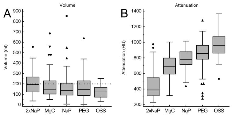
Figure 4.

Mean volume and attenuation of residual colonic fluid for each cathartic agent as quantified by automated volumetric QA tool. (A) Box and whisker plots showing mean residual colonic fluid volume. The mean residual volume was smallest when using the OSS regimen (125±60 ml) and significantly higher using 2xNaP regimen (206±125 ml) (p<0.0001), MgC regimen (184±125 ml) (p<0.01) and PEG regimen (166±114 ml) (p<0.05). NaP regimen showed also a higher residual volume (165±135 ml) than OSS regimen but without reaching a statistically difference (p=0.067). Note the outliers with high residual volumes with established cathartic agents that are not observed with OSS. Dotted line indicates 200 ml threshold of total residual colonic fluid. (B) Box and whisker plots showing the mean attenuation of the residual colonic fluid. The attenuation was significantly higher for the OSS regimen (955±167 HU) as compared to all of the other established cathartic agents, ranging from 406±191 HU for 2xNaP regimen (p<0.0001) to 843±193 HU for PEG regimen (p=0.012).
