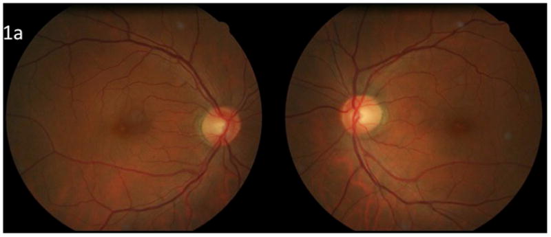
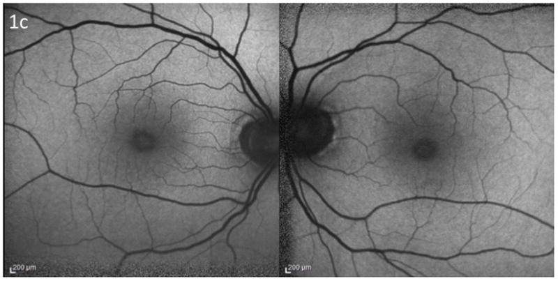
Figure 1.


Poppers maculopathy: a) Fundus photographs demonstrating bilateral foveal yellow spots b) Ocular coherence tomography of the macula demonstrating bilateral damage to the foveal photoreceptors c) Fundus autofluorescence photographs demonstrating central hyperautofluorescence. Courtesy of Dr. Catherine Vignal and Pr. Michel Paques, Paris, France.
