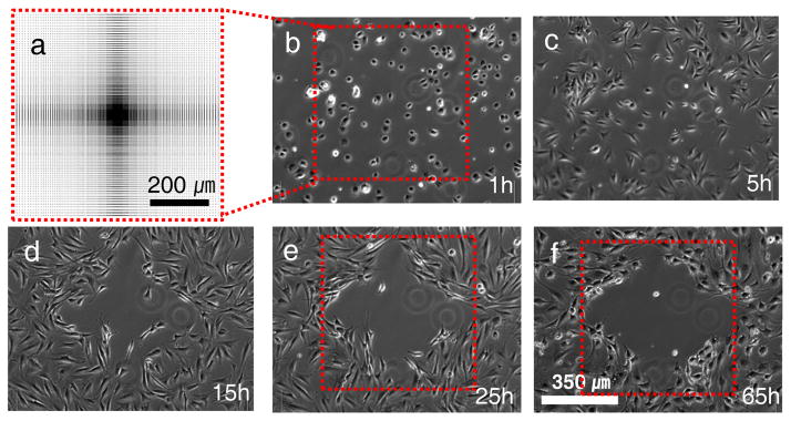
Figure 3.

Time-lapse phase-contrast images of NIH3T3 cells cultured on a patterned surface. a, Schematic diagram showing the full pattern design representing the pitch gradient. The distance between features varied between 1 and 10 μm, detailed dimensions are shown in Supplementary Fig. S5. The gradient was patterned on an area of 700 × 700 μm2 with crater diameter and depth of 1000 nm and 350 nm, respectively. b, At 1hr most cells are able to attach to the surface and spread even on the 1~2 μm spacing patterned area. c–f, However, the cells tend to migrate towards regions with greater pitch between the nanocraters. One day after cell seeding, clear boundary lines define a cell repellent region.
