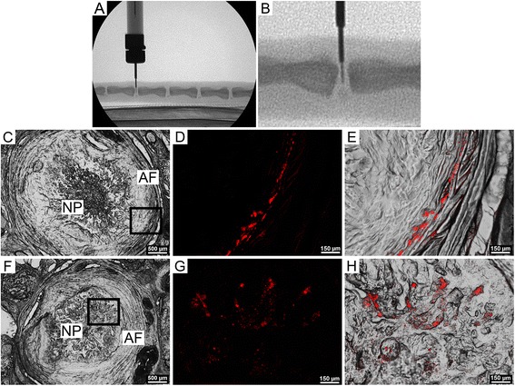
Figure 1.

In vivo delivery and localization microspheres. A. Delivery of microspheres to the rat NP from a custom 33-gauge needle under fluoroscopic guidance (lateral aspect). B. Higher magnification view of A. C. Differential interference contrast (DIC), axial image of entire disc 1 day post injection, showing NP and AF regions. D. Higher magnification view of the AF (inset from C.) showing microspheres (red). E. Microspheres (red, overlaying DIC image) embedded in the inner lamellae of the AF. F. Differential interference contrast, axial image of entire disc 14 days post injection, showing NP and AF regions. G. Higher magnification view (inset from F.) showing microspheres (red). H. Microspheres (red, overlaying DIC image), embedded in the NP.
